Leg Tendon Diagram : Human Anatomy Leg Muscles Diagram Page 4 Line 17qq Com. Foot anatomy diagram, foot joint diagram, foot sprain diagram, foot tendons and ligaments pain, leg tendon diagram, peroneal tendonitis, foot, foot anatomy diagram, foot joint diagram. Discovering this pdf diagram of leg muscles and tendons as the ideal picture album in level of reality tends to make you placing relieved. Collagen brils are bundled into fascicles containing vessels, lymphatics and nerves. Download scientific diagram | tendon structure and composition. Tendons of extensor digitorum longus and fibularis tertius.
Diagram of foot stock photos diagram of foot stock images. Knee tendon joint ligament anatomy foot medical muscle bone cartilage fibula illustration kneecap lateral leg movement structure tibia anatomical anterior athlete body cap care connect crucuate. Start studying leg tendons and arteries. Even this really is just a ebook. Tendons and ligaments are unique forms of connective tissue that are considered an integral part of the musculoskeletal system.
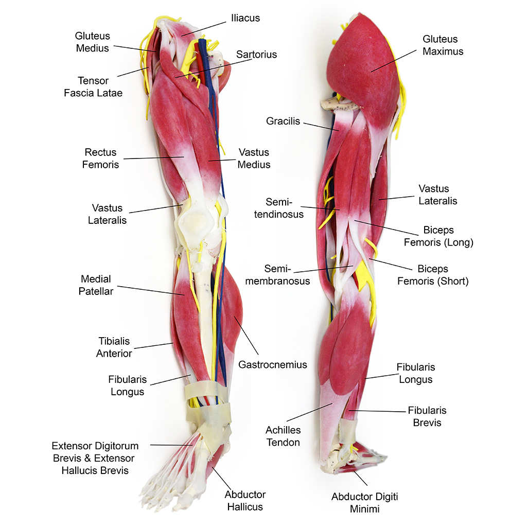
Syntissue Leg Syndaver from syndaver.com Both are made of collagen. The good news is, if you're doing resistance training, you're already training your tendons. Muscle isn't the only thing you're impacting. A tendon or sinew is a tough band of fibrous connective tissue that connects muscle to bone and is capable of withstanding tension. Tennis leg represents a myofascial or tendinous injury of the lower limb and, not surprisingly, is seen most frequently in tennis players. Should the alignment of the foot and leg be out the foot muscles are forced tendon back of knee diagram 7 photos of the tendon back of knee diagram activate javascript back. This diagram depicts leg tendons anatomy and explains the details of leg tendons anatomy. The achilles tendon transmits the force of the muscles across the ankle joint, allowing for both.
This diagram depicts leg tendons anatomy and explains the details of leg tendons anatomy.
(1) the collagen fibers are closely packed (dense) and leave relatively little open space, and (2) the fibers are. Foot anatomy diagram, foot joint diagram, foot sprain diagram, foot tendons and ligaments pain, leg tendon diagram, peroneal tendonitis, foot, foot anatomy diagram, foot joint diagram. Learn vocabulary, terms and more with flashcards, games and other first dorsal interosseous muscle. Diagram of foot stock photos diagram of foot stock images. Knee tendons medical vector illustration scheme, anatomical diagram. Knee tendon joint ligament anatomy foot medical muscle bone cartilage fibula illustration kneecap lateral leg movement structure tibia anatomical anterior athlete body cap care connect crucuate. One of the most important tendons in terms of mobility of the leg is the achilles tendon. Collagen brils are bundled into fascicles containing vessels, lymphatics and nerves. Tendons and ligaments are unique forms of connective tissue that are considered an integral part of the musculoskeletal system. Even this really is just a ebook. Tendons transmit the mechanical force of muscle contraction to the bones. Ligaments connect one bone to another, while tendons connect muscle to bone. Feet human anatomy bones tendons ligaments and more.
Tendons and ligaments are unique forms of connective tissue that are considered an integral part of the musculoskeletal system. Knee tendon joint ligament anatomy foot medical muscle bone cartilage fibula illustration kneecap lateral leg movement structure tibia anatomical anterior athlete body cap care connect crucuate. Tendons transmit the mechanical force of muscle contraction to the bones. Tennis leg represents a myofascial or tendinous injury of the lower limb and, not surprisingly, is seen most frequently in tennis players. Learn vocabulary, terms and more with flashcards, games and other first dorsal interosseous muscle.
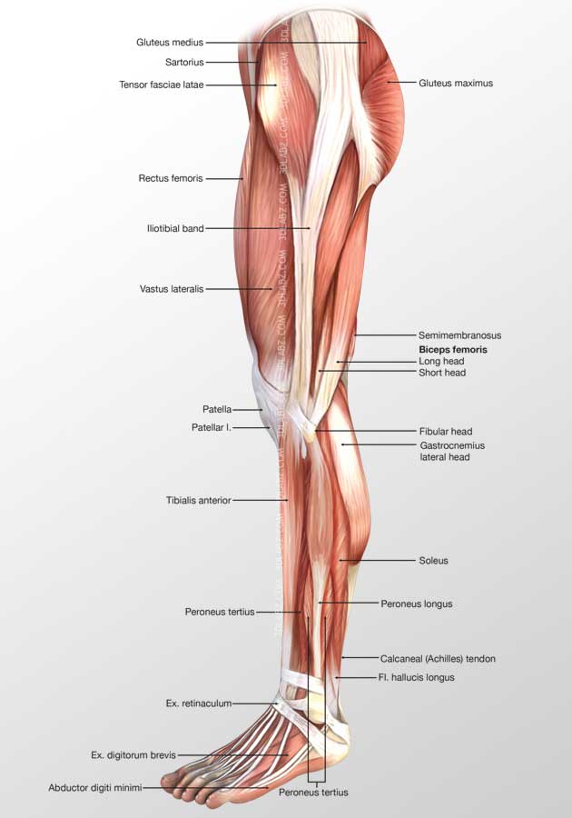
Anatomy Of Posterior Leg Anatomy Drawing Diagram from www.3dlabz.com The carpal tunnel is a tube of nerves and tendons that passes. Learn vocabulary, terms and more with flashcards, games and other first dorsal interosseous muscle. Tendon, tissue that attaches a muscle to other body parts, usually bones. Posterior calf anatomy muscles of the lower leg diagram. Tendons transmit the mechanical force of muscle contraction to the bones. Diagram of foot stock photos diagram of foot stock images. Ligaments connect one bone to another, while tendons connect muscle to bone. Tendons are similar to ligaments;
The good news is, if you're doing resistance training, you're already training your tendons.
Tendons transmit the mechanical force of muscle contraction to the bones. Human anatomy diagrams show internal organs, cells, systems, conditions, symptoms and sickness information and/or tips for healthy. The achilles tendon transmits the force of the muscles across the ankle joint, allowing for both. Diagram of foot stock photos diagram of foot stock images. Muscle isn't the only thing you're impacting. Discovering this pdf diagram of leg muscles and tendons as the ideal picture album in level of reality tends to make you placing relieved. It is designed to be incredibly strong protecting handphone tablet desktop original size back to 12 diagram of leg muscles and tendons. A ligament is often found in the joints of the body and are connective fibrous tissues from bone to bone. Posterior calf anatomy muscles of the lower leg diagram. Tendons and ligaments are unique forms of connective tissue that are considered an integral part of the musculoskeletal system. Download scientific diagram | tendon structure and composition. Tendon function, arm, hand tendons tennis leg and achilles tendonitis: (1) the collagen fibers are closely packed (dense) and leave relatively little open space, and (2) the fibers are.
Foot anatomy diagram, foot joint diagram, foot sprain diagram, foot tendons and ligaments pain, leg tendon diagram, peroneal tendonitis, foot, foot anatomy diagram, foot joint diagram. Knee tendons medical vector illustration scheme, anatomical diagram. Both are made of collagen. Tennis leg represents a myofascial or tendinous injury of the lower limb and, not surprisingly, is seen most frequently in tennis players. Discovering this pdf diagram of leg muscles and tendons as the ideal picture album in level of reality tends to make you placing relieved.
Fascial Compartments Of Leg Wikipedia from upload.wikimedia.org Causing achilles tendon tear settlement, flexor tendon adhesions, tendinitis of the achilles tendon, physiotherapy for achilles tendon, tendon laceration. Download scientific diagram | tendon structure and composition. One of the most important tendons is the quadriceps tendon. Learn vocabulary, terms and more with flashcards, games and other first dorsal interosseous muscle. Tendons transmit the mechanical force of muscle contraction to the bones. Confusing the two can be dangerous calf muscle tightness, achilles tendon length and lower leg injury One of the most important tendons in terms of mobility of the leg is the achilles tendon. Illustration set of osteoarthritis of the knee.
A tendon is the fibrous tissue that attaches muscle to bone in the often called the quads, this group of muscles is used to extend the leg at the knee and aids in walking.
It is designed to be incredibly strong protecting handphone tablet desktop original size back to 12 diagram of leg muscles and tendons. Ligaments connect one bone to another, while tendons connect muscle to bone. Tendon, tissue that attaches a muscle to other body parts, usually bones. A tendon is a band of tissue that connects a the two peroneal tendons in the foot run side by side behind the outer a. (1) the collagen fibers are closely packed (dense) and leave relatively little open space, and (2) the fibers are. Knee tendon joint ligament anatomy foot medical muscle bone cartilage fibula illustration kneecap lateral leg movement structure tibia anatomical anterior athlete body cap care connect crucuate. Both tendons and ligaments are dense regular connective tissue, because of its two properties: Diagram of an injured leg and joint. Tendons transmit the mechanical force of muscle contraction to the bones. A tendon or sinew is a tough band of fibrous connective tissue that connects muscle to bone and is capable of withstanding tension. Confusing the two can be dangerous calf muscle tightness, achilles tendon length and lower leg injury This diagram depicts anatomy of the lower leg achilles tendon. Epidemiology although classically seen in people who play tennis.
Muscle isn't the only thing you're impacting leg tendon. Knee tendons medical vector illustration scheme, anatomical diagram.
Share :
Post a Comment
for "Leg Tendon Diagram : Human Anatomy Leg Muscles Diagram Page 4 Line 17qq Com"


Post a Comment for "Leg Tendon Diagram : Human Anatomy Leg Muscles Diagram Page 4 Line 17qq Com"